Showing posts with label ANATOMY. Show all posts

Monday, October 17, 2011

Preauricular Incision For TMJ surgery


General considerationsThe preauricular approach can be used to access and treat fractures in the mandibular condylar head and neck region. Many surgeons perform temporal mandibular joint (TMJ) surgery and routinely use this incision to access the superior portion of the mandibular condylar process.
The illustration demonstrates the access and the amount of exposure.
Neurovascular structures
Branches of the facial nerve may be involved in this incision and dissection.
The superficial temporal artery and vein are commonly encountered in this surgical approach. The vessels should be conserved if possible.
Exposure offered by extraoral approachesSubmandibular approach
Retromandibular
  • Transparotid
  • Retroparotid

Preauricular approach
Facelift incision (rhytidectomy)
Skin incision
General consideration
Use of a solution containing vasoconstrictors ensures hemostasis at the surgical site. The two options currently available are the use of local anesthetic or a physiologic solution with vasoconstrictor alone.
Use of a local anesthetic with vasoconstrictor may impair the function of the facial nerve and impede the use of a nerve stimulator during the surgical procedure. Therefore, consideration should be given to using a physiological solution with vasoconstrictor alone or injecting the local anesthetic with vasoconstrictor very superficially.
Make the incision in a preauricular skin crease.
Dissection
Locating temporalis fascia
Carry the incision through the skin and subcutaneous tissues to the depth of the temporalis fascia. The temporalis fascia is a glistening white tissue layer that is best appreciated in the superior portion of the incision.
The superficial temporal vessels may be retracted anteriorly with the skin flap (sectioning some posterior and superior branches) or left in place (sectioning frontal branches).
The zygomatic arch can easily be palpated at this point of the dissection. The lateral pole of the mandibular condyle can also be palpated. This can be facilitated by having a surgical assistant manipulate the jaw.
Incising temporalis fasciaMake an oblique incision parallel to the frontal branch of the facial nerve, through the superficial layer of the temporalis fascia above the zygomatic arch.
Dissection of the joint capsuleInsert the periosteal elevator beneath the superficial layer of the temporalis fascia and strip the periosteum off the lateral zygomatic arch.
Dissection will be carried inferiorly to expose the capsule of the TMJ.
Coronal view of dissection to the lateral portion of the zygomatic arch and mandibular condyle region.
Note: the frontal branch of the facial nerve is protected within the superficial layer of the deep temporalis fascia.
Optional: capsule incision
In the rare case of treating condylar head fractures the TMJ capsule is incised in an open manner.
Dissection can be carried inferiorly in a subperiosteal plane to reach the neck of the mandibular condyle.
A disadvantage of this approach is that the surgeon can reach only a limited portion of the condylar neck region.
Wound closure
If the TMJ capsule has been incised to access the condylar head it must be closed as the first step.
The temporalis fascia is closed as the next step.
Skin and subcutaneous sutures are placed.
A pressure dressing may be placed over this wound according to surgeon’s preference.
Friday, October 7, 2011

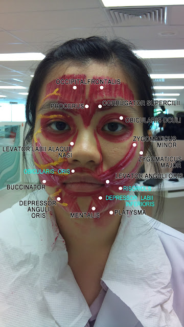
Muscles of Facial Expression & Facial Nerve (Clinical Session)


Muscles of Facial Expression

Motor Branch of Facial Nerve


Temporal Branch innervates :
  • Frontalis
  • Obicularis Oculi
  • Corrugator Supercilii
  • Anterior & Superior Auricular Muscles


Zygomatic Branch innervates :
  • Obicularis Oris


Buccal Branch innervates :
  • Buccal region

Mandibular Branch innervates :
  • Muscles of Lower Lip
  • Chin

Cervical Branch innervates :
  • Platysma

Trigeminal nerve and venous drainage of face


TRIGEMINAL NERVE (CNV)
Branches and Routes of the Maxillary and Mandibular Divisions

VENOUS DRAINAGE OF HEAD AND NECK
  1. Supratrochlear and Supraorbital veins form Angular vein.
  2. Angular vein is an upper tributary of the Facial vein.
  3. Superficial Temporal vein and Maxillary vein from Paroitd gland unite to form the Retromandibular vein.
  4. Posterior Auricular vein drains into Retromandibular vein.
  5. Retromandibular vein divides into two branches; Anterior and Posterior branch.
  6. Anterior branch of Retromandibular vein joins anterior Facial vein anteriorly.
  7. Posterior branch of Retromandiular vein joins the External Jugular vein posteriorly.
  8. Occipital vein drains into Suboccipital Venous Plexus in Suboccipital Triangle.
  9. There are emissary veins connecting deep veins to superficial veins.
           > Facial vein - Deep Facial vein
           > Intercranial Cavernous Sinus
           > Pharyngeal Venous Plexus

Friday, August 12, 2011

MUSCLES OF TONGUE


  • DIVIDED INTO TWO TYPES OF MUSCLES- EXTRINSIC & INTRINSIC
  • EXTRINSIC MUSCLES - 
  1. GENIOGLOSSUS- origin: Genial tubercle of mandible; insertion: Tip and dorsum of tongue & hyoid bone.
  2. HYOGLOSSUS-origin: Greater cornua of hyoid bone; insertion: Side of tongue 
  3. STYLOGLOSSUS- origin: styloid process; insertion: Side of tongue
  4. PALATOGLOSSUS-origin: palatine aponeurosis; insertion: side of the tongue at the junction of tongue and pharynx.
  • INTRINSIC MUSCLES-
  1. SUPERIOR LONGITUDNAL
  2. INFERIOR LONGITUDNAL
  3. TRANSVERSE
  4. VERTICAL
  • NERVE SUPPLY OF TONGUE-
  1. SENSORY NERVE SUPPLY- Lingual nerve branch of mandibular nerve(general nerve for sensation) & chorda tympani nerve branch of facial nerve(Taste sensation except circumvallate papillae) supply anterior two third of tongue.Glossopharyngeal nerve supplies sensory & taste  innervation to posterior one third of tongue including circumvallate papillae.Laryngeal branch of Vagus nerve supply most posterior part of tongue
  2. MOTOR NERVE SUPPLY-Hypoglossal nerve to all muscles except palatoglossus which is supplied by cranial part of accessory nerve.
  • PAPILLAE- A small, round or cone-shaped bump on the surface of the tongue. There are several types of papillae in the mouth, and all but one type containtaste buds.
    Fungiform - taste bud-containing papillae located on the front two-thirds of the tongue. They can be seen as red bumps (the bumps that stand out in contrast to the pinkness of the rest of your tongue); under magnification, they look a bit like mushrooms (fungi).
    Circumvallate - taste bud-containing papillae toward the very back of the tongue; they are placed in an inverted “V.” It can be very hard to see your own, but it is fairly easy to see these in another person, especially if you use a flashlight.
    Foliate - taste bud-containing papillae located very far back on the sides of the tongue; they look like a series of folds or lines and can be very difficult to see.
    Filiform - papillae that do not contain taste buds. They cover the surface of the tongue in great abundance and are largely responsible for the texture of the tongue. The only purpose it serves in tasting is that it can help to hold taste compounds on the tongue, increasing the chance that the taste compound will interact with a taste receptor cell.

Thursday, August 11, 2011


The Suprahyoid Muscles
  • This group of muscles is located superior to the hyoid bone and connects to this bone and the skull.
  • This group includes the mylohyoid, geniohyoid, stylohyoid and digastric muscles.

The Mylohyoid Muscles 
  • These are thin, flat triangular muscles that form a sling inferior to the tongue.
  • They form the floor of the mouth.
  • Role of these muscles in grinding the food in the mouth.
  • Superior attachment: mylohyoid line of mandible.
  • Inferior attachment: raphe and body of hyoid bone.
  • Innervation: mylohyoid nerve, a branch of the inferior alveolar nerve.
  • It elevates the hyoid bone, floor of the mouth and the tongue during swallowing and speaking.

Geniohyoid Muscles 
  • These are short narrow muscles that contact each other in the median plane.
  • They are located superior to the mylohyoid muscles, where they reinforce the floor of the mouth.
  • Superior attachment: inferior mental spine of mandible.
  • Inferior attachment: body of hyoid bone.
  • Innervation: C1 via the hypoglossal nerve (CN XII).
  • It pulls the hyoid bone anterosuperiorly, and shortens the floor of the mouth and widens the pharynx.

The Stylohyoid Muscles 
  • These muscles form a small slip on each side, which is nearly parallel to the posterior belly of the digastric muscle.
  • Superior attachment: styloid process of the temporal bone.
  • Inferior attachment: body of hyoid bone.
  • Innervation: Stylohyoid branch of facial nerve (CN VII).
  • It elevates and retracts the hyoid bone, thereby elongating the floor of the mouth.

The Digastric Muscles 

  • Each of the strap-like muscles has two bellies (G. gaster, belly) that descend toward the hyoid bone.
  • They are joined by an intermediate tendon that is connected to the body and the greater horn of the hyoid bone by a strong loop or sling of fibrous connective tissue.
  • This fibrous pulley allows the intermediate tendon to slide anteriorly and posteriorly.
  • Superior attachment: anterior belly-digastric fossa of mandible, posterior belly-mastoid notch of temporal bone.
  • Inferior attachment: intermediate tendon to body and greater horn of hyoid bone.
  • Innervation: anterior belly-mylohyoid nerve, a branch of the inferior alveolar nerveposterior belly-facial nerve (CN VII).
  • It depresses the mandible and raises the hyoid bone. Also, it steadies the hyoid bone during swallowing and speaking.


Mylohyoid muscle alnwith lingual nerve and submandibular and sunlingual gland 

HYOID BONE


hyoid bone-


hyoid bone
Location of the hyoid

hyoid anterosuperior aspect
Anterosuperior aspect

hyoid right aspect
Right aspect

A small, U-shaped bone situated centrally in the upper part of the neck, beneath the mandible but above the larynx near the level of the third cervical vertebra. It can be felt by pressing one's finger into the crease where the chin becomes the neck. The hyoid bone consists of three separate parts – the body, and the left and right greater and lesser cornu (horns) – which fuse in early adulthood.

The function of the hyoid is to provide an anchor point for the muscles of the tongue and for those in the upper part of the front of the neck.

The hyoid is (uniquely in the vertebrate skeleton) not joined to any other bone but is suspended in position by muscles that connect it to the mandible, to the styloid process of each temporal bone at the base of the skull, to the thyroid cartilage, to the sternum, and to the scapula. The important muscles that are attached to the hyoid bone are shown in the lower two diagrams.